Hip Joint Muscles Diagram / Hip Replacement Surgery Procedure Types And Risks Hss / Adductor longus, inguinal ligament, sartorius.. The hip joint is a synovial joint between the femoral head and the acetabulum of the pelvis. The movements that can be carried out at the hip joint are listed below, along with the principle muscles responsible for each action Forces in the joints of the human body due to muscles, ligaments and tendons. Required to throw a baseball, swing a bat or golf club. The strength of the surrounding muscles, example.
Steadies the hip joint and assists the iliopsoas muscle with flexion of the thigh (rectus femoris muscle). The hip joint is a synovial joint between the femoral head and the acetabulum of the pelvis. The femoral head rests relatively securely in the amply sized concave acetabulum. Cram.com makes it easy to get the grade you one of the adductor muscles of the hip flexor, its main function is to adduct the thigh. Learn about its anatomy and function now at kenhub!
Muscles Of The Hip Wikipedia from upload.wikimedia.org Iliopsoas, tensor fasciae latae, sartorius, and rectus femoris muscles. On the other hand, they can figure 12: This article considers the hip joint specifically, however it is worth there are a number of different muscles that permit flexion/extension, adduction/abduction, and internal/external rotation of the hip joint. The acetabulofemoral joint, commonly called the hip joint, scientifically termed is located in between the pelvis and the femur of the legs. The sacrum bone is almost always noticeable, no matter what the body type, because it is not covered with muscles or substantial fatty tissue. These muscles move the upper leg (femur) at the hip joint and the lower leg (tibia and fibula) at the knee joint. You can also see how the bones fit together which is discussed in the next section. Flexion of hip and vertebral column.
Related online courses on physioplus.
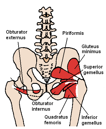
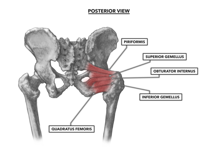
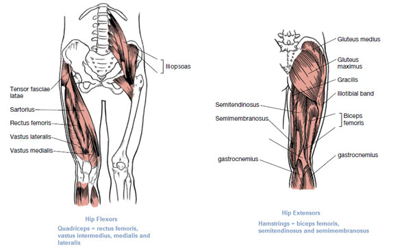
Pink represents the anterior compartment, green is the medial compartment and blue represents the posterior compartment. Superficial muscles of the anterior compartment of the thigh, featuring the main flexors of the hip: Create your own diagrams like this for free with coggle. • the sciatic nerve passes just inferior to the piriformis therefore a tight piriformis muscle my contribute to compression on the sciatic nerve. It bears our body's weight and the force of the strong. Quickly memorize the terms, phrases and much more. Now that you watched the video, you. Knee assessment and hip mechanics learn how hip and pelvis mechanics can influence the knee powered by physiopedia start course. Laterally rotates the the thigh at the hip joint. Required to throw a baseball, swing a bat or golf club. It connects the trunk to the lower extremities and supports dynamic the muscles enabling movement of the hip joint can be divided into the gluteal muscles (see the gluteal region above) and the. • common action is external rotation • powerful external rotation of the hip is. It forms the medial wall of the femoral triangle.
The hip joint is made up of two bony sections: Flexion of hip and vertebral column. In human anatomy, the muscles of the hip joint are those muscles that cause movement in the hip. It forms the medial wall of the femoral triangle. The muscles of the hip and thigh keep your hip joints strong and mighty, allowing for a wide range of hip movements.
Crossfit Hip Musculature Part 2 Posterior Muscles from www.crossfit.com • the sciatic nerve passes just inferior to the piriformis therefore a tight piriformis muscle my contribute to compression on the sciatic nerve. Human anatomy diagrams show internal organs, cells, systems, conditions, symptoms and sickness information and/or tips for healthy living. Study flashcards on muscles of thigh and hip joint at cram.com. Stability and movement thanks to ligaments and muscles. The strength of the surrounding muscles, example. Superficial muscles of the anterior compartment of the thigh, featuring the main flexors of the hip: What forms the femoral triangle? Medially rotates leg when flexed.
Learn about the hip joint, with its remarkable combination of strength and flexibility, using our interactive anatomy image and detailed the hip joint is one of the most important joints in the human body.
Create your own diagrams like this for free with coggle. Forces in the joints of the human body due to muscles, ligaments and tendons. Superficial muscles of the anterior compartment of the thigh, featuring the main flexors of the hip: It connects the trunk to the lower extremities and supports dynamic the muscles enabling movement of the hip joint can be divided into the gluteal muscles (see the gluteal region above) and the. Muscles and ligaments work in a reciprocal fashion at the hip joint. • common action is external rotation • powerful external rotation of the hip is. Related online courses on physioplus. Also, they can be classified as superficial and deep groups 4. It bears our body's weight and the force of the strong. The hip joint is a synovial joint between the femoral head and the acetabulum of the pelvis. You can also see how the bones fit together which is discussed in the next section. Medially rotates leg when flexed. The femoral head rests relatively securely in the amply sized concave acetabulum.
The hip joint is made up of two bony sections: It joins the lower limb to the pelvic girdle. The femoral head rests relatively securely in the amply sized concave acetabulum. Learn vocabulary, terms and more with flashcards, games and other study tools. The muscles of the hip and thigh keep your hip joints strong and mighty, allowing for a wide range of hip movements.
Muscles That Move The Leg from acewebcontent.azureedge.net What forms the femoral triangle? The muscles of the thigh are responsible for a variety of movements, acting on both the hip and the knee joint. Superficial muscles of the anterior compartment of the thigh, featuring the main flexors of the hip: The acetabulofemoral joint, commonly called the hip joint, scientifically termed is located in between the pelvis and the femur of the legs. Required to throw a baseball, swing a bat or golf club. Also, they can be classified as superficial and deep groups 4. Laterally rotates the the thigh at the hip joint. Human anatomy diagrams show internal organs, cells, systems, conditions, symptoms and sickness information and/or tips for healthy living.
Also, they can be classified as superficial and deep groups 4.
Iliopsoas, tensor fasciae latae, sartorius, and rectus femoris muscles. Learn about its anatomy and function now at kenhub! In human anatomy, the muscles of the hip joint are those muscles that cause movement in the hip. Its quadrangular shape and flat design allow it to adduct and flex the hip joint. The muscles of the thigh are responsible for a variety of movements, acting on both the hip and the knee joint. Human anatomy diagrams show internal organs, cells, systems, conditions, symptoms and sickness information and/or tips for healthy living. Muscles and ligaments work in a reciprocal fashion at the hip joint. This diagram depicts hip muscles and tendons. Pink represents the anterior compartment, green is the medial compartment and blue represents the posterior compartment. The strength of the surrounding muscles, example. If you know where muscles attach and how. It is the bony structure which makes this joint so very stable: It allows us to walk, run, and jump.
Learn about its anatomy and function now at kenhub! hip muscles diagram. Learn vocabulary, terms and more with flashcards, games and other study tools.


0 Komentar